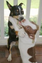
HEALTH TESTING : All of our dogs are tested and registered through OFA, and almost all our dogs are in the process of finishing their CHIC numbers with AKC. All results are posted on our website below each dogs information. Please ask if you have any questions.
1. Primary Lens Luxation in Teddy Roosevelt Terriers (PLL)
Teddy Roosevelt Terrier breeders and Rat Terriers owners need to be aware of an eye issue that was recently discovered in this Terrier breed. It is an issue that has lurked unknowingly in the breed for many years. The good news is, as of October 2010, an invaluable “test” became available to find the DNA marker for Primary Lens Luxation. When selecting a Teddy Roosevelt Terrier or Rat Terrier , ask for PROOF of their testing results. If the dogs’ parents have not been tested or proof is withheld … ask for this testing to be done prior to purchase.
WHAT IS PLL? Primary Lens Luxation is the dislocation or displacement of the lens within the eye. The lens is the clear structure in the eye, consisting of two rounded or convex surfaces, that focuses light rays to form an image onto the retina. Normally the lens is suspended between the iris (the colored portion of the eye) and the vitreous (the clear gel in the back of the eye), and is held in place by small fibers called zonules or suspensory ligaments. Should the zonules break, the lens can either become partially dislocated (subluxated) from its normal position or completely dislocated (luxated). When the lens detaches and falls forward into the anterior chamber in front of the pupil, it is called an Anterior Luxation. When it falls back into the rear portion of the eye, it is called a Posterior Luxation.
HOW DOES A DOG GET IT? Primary Lens Luxation is an inherited disorder in which the zonules or suspensory fibers degenerate. The condition occurs mainly in the terrier breeds, namely the Parson Russell Terrier, Tibetan Terrier, Smooth Fox Terrier and Rat Terrier. Primary luxations are also seen in the Border Collie, the Australian Cattle dog (blue heeler), and sporadically in other breeds. Although the underlying reasons for the lens luxation are not well understood, inflammation or a defect in the zonules may play a role. With primary lens luxation, both eyes are prone to dislocation of the lens.
HOW CAN I TEST MY DOG? Before October 10, 2010 there was NO test for PLL. Through the partnership of the University of Missouri College of Veterinary Medicine and OFA (Orthopedic Foundation for Animals), there is NOW a DNA test for this mutation. The cost is $65.
HOW IS THE TEST PERFORMED? A Test Kit will include a cheek swab and DNA marker card with instructions how to perform this simple test. The inside of the dog’s mouth/cheek is swabbed to collect cells and saliva. Then the saliva is spread onto the card. The card is sent to OFA.
WHAT WILL THE TEST REVEAL? The DNA test results will categorize dogs as follows: Affected, Carrier, or Normal/Clear
AFFECTED: This finding indicates that TWO copies of the disease gene are present in the dog. Unfortunately, the dog will be medically Affected by the disease. Appropriate treatment should be pursued by consulting a veterinarian. An AFFECTED dog will have 2 mutated copies of the gene. The vast majority of these dogs will luxate at 4-8yrs of age, the typical age of onset for PLL. There were a few dogs in the study group that tested as AFFECTED but did not luxate until after 8 yrs of age, and some dogs testing AFFECTED have died from other causes without luxating. A search of published veterinary literature revealed that about 10% of the dogs reported to be clinically Affected with PLL had onset of symptoms after 8 yrs of age. Because of this, the test results will say “AFFECTED/HIGH RISK.
CARRIER: This finding indicates that ONE copy of the disease gene is present in your dog, but that it will NOT exhibit disease symptoms. Carriers will NOT have medical problems as a result. Dogs with Carrier status can be enjoyed without the fear of developing medical problems but will pass on the disease gene 50% of the time. Carriers have one normal and one mutated copy of the gene. They could pass either the normal copy or the mutated copy on to their offspring. Because there were very few cases of dogs in the research groups testing CARRIER who did appear to have PLL, the test results will say “CARRIER/LOW RISK.
NORMAL: This finding indicates that the GENE IS NOT PRESENT in your dog. Therefore, when used for breeding, a Clear dog will NOT pass on the disease gene. A dog testing NORMAL has 2 normal copies of the gene, is not at risk for developing PLL, and can only pass a normal copy of the gene to any offspring. Some sites use the word Normal or Clear. Both these words mean the same thing. It means that the dog in question is NOT a Carrier nor is it Affected.
CLEAR BY PARENTAGE (CBP): You may see health clearances with the wording "Cleared By Parentage" or "CBP" on breeder sites. This means both sire and dam are proven Normal/Clear and that pair bred together can never produce an Affected or a Carrier. Some sites use the word Normal or Clear. Both these words mean the same thing. It means that the dog in question is NOT a Carrier nor is it Affected. Always request to see parent testing results for clear Sire and Dam.
|
2. Patellas
THE LUXATING KNEE
What is Patellar Luxation?
The patella, or kneecap, is part of the stifle joint (knee). In patellar luxation, the kneecap luxates, or pops out of place, either in a medial or lateral position.
Bilateral involvement is most common, but unilateral is not uncommon. Animals can be affected by the time they are 8 weeks of age. The most notable finding is a knock-knee (genu valgum) stance. The patella is usually reducible, and laxity of the medial collateral ligament may be evident. The medial retinacular tissues of the stifle joint are often thickened, and the foot can be seen to twist laterally as weight is placed on the limb.
Patellar Luxation Categories
Patellar luxations fall into several categories:
- Medial luxation; toy, miniature, and large breeds
- Lateral luxation; toy and miniature breeds
- Lateral luxation; large and giant breeds.
- Luxation resulting from trauma; various breeds, of no importance to the certification process.
3. Hips
THE DYSPLASTIC HIP JOINT
 Hip Dysplasia is a terrible genetic disease because of the various degrees of arthritis (also called degenerative joint disease, arthrosis, osteoarthrosis) it can eventually produce, leading to pain and debilitation.
The very first step in the development of arthritis is articular cartilage (the type of cartilage lining the joint) damage due to the inherited bad biomechanics of an abnormally developed hip joint. Traumatic articular fracture through the joint surface is another way cartilage is damaged. With cartilage damage, lots of degradative enzymes are released into the joint. These enzymes degrade and decrease the synthesis of important constituent molecules that form hyaline cartilage called proteoglycans. This causes the cartilage to lose its thickness and elasticity, which are important in absorbing mechanical loads placed across the joint during movement. Eventually, more debris and enzymes spill into the joint fluid and destroy molecules called glycosaminoglycan and hyaluronate which are important precursors that form the cartilage proteoglycans. The joint's lubrication and ability to block inflammatory cells are lost and the debris-tainted joint fluid loses its ability to properly nourish the cartilage through impairment of nutrient-waste exchange across the joint cartilage cells. The damage then spreads to the synovial membrane lining the joint capsule and more degradative enzymes and inflammatory cells stream into the joint. Full thickness loss of cartilage allows the synovial fluid to contact nerve endings in the subchondral bone, resulting in pain. In an attempt to stabilize the joint to decrease the pain, the animal's body produces new bone at the edges of the joint surface, joint capsule, ligament and muscle attachments (bone spurs). The joint capsule also eventually thickens and the joint's range of motion decreases.
No one can predict when or even if a dysplastic dog will start showing clinical signs of lameness due to pain. There are multiple environmental factors such as caloric intake, level of exercise, and weather that can affect the severity of clinical signs and phenotypic expression (radiographic changes). There is no rhyme or reason to the severity of radiographic changes correlated with the clinical findings. There are a number of dysplastic dogs with severe arthritis that run, jump, and play as if nothing is wrong and some dogs with barely any arthritic radiographic changes that are severely lame.
4. Heart
CONGENITAL CARDIAC DISEASE AND THE OFA
Congenital heart diseases in dogs are malformations of the heart or great vessels. The lesions characterizing congenital heart defects are present at birth and may develop more fully during perinatal and growth periods. Many congenital heart defects are thought to be genetically transmitted from parents to offspring; however, the exact modes of inheritance have not been precisely determined for all cardiovascular malformations.
Developmental Inherited Cardiac Diseases (SAS and Cardiomyopathy)
At this time inherited, developmental cardiac diseases like subaortic stenosis and cardiomyopathies are difficult to monitor since there is no clear cut distinction between normal and abnormal. The OFA will modify the congenital cardiac database when a proven diagnostic modality and normal parameters by breed are established. However at this time, the OFA cardiac database should not be considered as a screening tool for these diseases.
5. Canine degenerative myelopathy, also known as chronic degenerative radiculomyelopathy, is an incurable, progressive disease of the canine spinal cord that is similar in many ways to amyotrophic lateral sclerosis. Onset is typically after the age of 7 years and it is seen most frequently in the German shepherd dog, Pembroke Welsh corgi, and boxer dog, though the disorder is strongly associated with a gene mutation in SOD1 that has been found in 43 breeds as of 2008, including the wire fox terrier, Chesapeake Bay retriever, Rhodesian ridgeback, and Cardigan Welsh corgi.[1][2] Progressive weakness and incoordination of the rear limbs are often the first signs seen in affected dogs, with progression over time to complete paralysis. Myelin is an insulating sheath around neurons in the spinal cord. One proposed cause of degenerative myelopathy is that the immune system attacks this sheath, breaking it down. This results in a loss of communication between nerves in lower body of the animal and the brain.